Two-photon microscopy and its application to skin imaging
Nov. 01, 2023
Traditional dermatopathologic testing usually involves cutting a certain size of skin tissue to make a pathology section, which is processed by HE staining and then viewed with microscopic imaging. This imaging method is invasive and time-consuming, so a large number of researchers are committed to developing non-invasive, in vivo, real-time, label-free imaging techniques. One such technique is two-photon microscopy. This is an imaging technique that combines the principles of two-photon absorption and excitation with laser scanning confocal microscopic imaging. It has become a very important imaging technique in the biomedical field since its introduction because of its large penetration depth, strong chromatographic imaging capability, excitation of exogenous and endogenous fluorophores, and submicron resolution.
01 Principles, methods and equipment
1.1 Two-photon imaging principle
Two-photon microimaging uses a micro objective to focus a near-infrared laser pulse on the order of a hundred femtoseconds into the sample, where the fluorescent molecules at the focal point undergo a two-photon absorption and jump to a higher energy level, emitting fluorescent photons as they return to the ground state. Under the control of the scanning system, the laser scans the sample in three axes, X,Y,Z, and records the gray value information of each pixel point, thus realizing 3D chromatographic imaging. In addition to the two-photon fluorescence process described above, other nonlinear effects occur during the imaging process. Multimodal multiphoton microimaging can be realized by choosing appropriate laser parameters and adjusting the corresponding detection bands.
1.2 Structure of the skin and distribution of endogenous fluorophores
The skin is the largest organ of the body. The human epidermis, except for the palm of the hand, can be divided into the stratum corneum, stratum granulosum, stratum basale, and then the dermis. The stratum corneum consists of the nucleus and organelles disappearing from the keratinocyte nucleus intercellular matrix, rich in keratin, with a strong TPEF signal; granular layer consists of 1~3 layers of flat or rhombic cells, cytoplasmic interior by a small number of mitochondria, melanin particles, such as fluorescent clusters, the TPEF signal is very weak. The basal layer is a layer of columnar or cuboidal basal cells, the nucleus is usually arranged above the cap-shaped melanin complex, with a strong TPEF signal.
1.3 Instrument Design
The handheld two-photon microscope is shown in Fig. 1. In the probe, the laser passes through two mirrors and is incident on a microelectromechanical scanning galvanometer, which is a MEMS scanning galvanometer that supports a frame rate of @512 × 512 pixels in a raster scanning mode, with a scanning field of view size of ~140 μm × 140 μm. The axial motion of the entire probe is controlled by a linear displacement stage, which allows for layer-slicing scans of samples with thicknesses of more than 300 μm. The femtosecond laser is focused onto the sample by an objective lens with a numerical aperture of 0.9. The signal generated by the sample excitation is collected in a glass fiber bundle and transmitted along the cable between the main unit and the handle back to the light probe module in the main unit. The module consists of a TPEF channel (420~580 nm) and a SHG channel.
The two-photon fluorescence signal and the second harmonic signal were detected separately. The signal intensity of each pixel is stored as a 16-bit grayscale value and processed by the control software based on the ZYNQ main control board and host computer to reconstruct the scanned image. The microscope used an objective lens with a numerical aperture of 0.9 immersion medium of silicone oil or silicone gel to achieve a lateral resolution of less than 0.6 μm and an axial resolution of less than 3.0 μm.
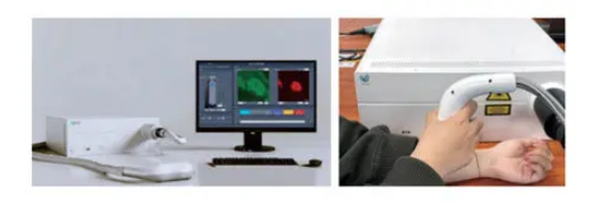
The two-photon microscope can reconstruct the 3D structure of the skin in the measured area by scanning the image layer by layer, and obtain the three-dimensional topographical features of the skin, such as the thickness of the stratum corneum and the thickness of the epidermis. The fluorescence channel can clearly distinguish the cytoplasm and nucleus, and observe the dispersion and clustering of mitochondria, the density of elastic fibers and the composition of the network, etc.; the second harmonic channel can observe the distribution of collagen fibers; through the presence or absence of collagen, it can be judged whether the imaging depth reaches the true epidermal junction, which is convenient for the accurate localization of the basal layer. On the other hand, since the two-photon fluorescence signal originates from the fluorescence of molecules, and its signal intensity is proportional to the concentration of the luminescent substance, two-photon microscopy can also be used to carry out functional imaging, such as in vivo detection of the oxygen consumption rate of keratinocytes, measurement of the skin's SAAID/ELCOR index, and the detection of fluorescence rise rate and fall rate during the process of arterial blockage and refocusing, and so on.
Handheld two-photon microscopes are used for much more than skin imaging. As a general-purpose two-photon microscope, it can be utilized to observe isolated samples, biopsy animals and plants, and directly observe cultured cells and microorganisms. In addition, using the same handheld probe technology, it can be a handheld two-photon microscope of the appropriate wavelength for observing a variety of endogenous and exogenous fluorescence and high harmonic generation simply by developing a miniaturized femtosecond laser of the appropriate wavelength.
Portable, handheld two-photon microscopes allow for the realization of technology that was previously only available in benchtop two-photon microscopes or removable benchtop two-photon microscopes again. Weighing only 12kg, the development of handheld two-photon microscope, for the majority of skin-related research workers, doctors, beauty and cosmetics industry practitioners to provide a convenient choice, can be realized anytime, anywhere in the body, in situ, non-invasive, non-labeling high temporal and spatial resolution two-photon microscopic imaging, in the field of skin disease diagnostic aids, cosmetic efficacy testing, medical and beauty assessment has a broad application prospects.
CLZ Optical Co., Ltd.has been manufacturing and trading optical components for many years such as optical domes, optical prisms, optical windows and so on, we also could provide OEM service, customized for our customer, please contact us free time if you have any needs!
Previous: Manufacture of optical components


